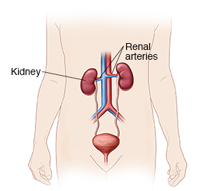
Renal Angiography
Renal angiography is an imaging test used to study the blood vessels in your kidneys. The procedure is done through a thin, flexible tube called a catheter. The catheter is put into a blood vessel through a small cut or incision. X-ray dye, also called contrast medium, is injected. The dye makes the blood vessels easier to see on X-ray images. X-rays are then taken. The procedure is often done by a specially trained healthcare provider called an interventional radiologist. This is a healthcare provider who is certified by the American Board of Radiology to use minimally invasive image-guided procedures to diagnose and treat diseases.
Before your procedure
Follow any directions you are given on how to get ready. This includes any directions you're given for not eating or drinking before the procedure.
Tell your healthcare provider if you are:
-
Allergic to X-ray dye or other medicines
-
Are breastfeeding
-
Are pregnant or think you may be pregnant
-
Have any medical conditions
-
Have had any recent illnesses
Tell your healthcare provider about all medicines you take. You may need to stop taking some or all of them before the test. This includes:
-
All prescription medicines
-
Herbs, vitamins, and other supplements
-
Over-the-counter medicines, such as aspirin or ibuprofen
-
Illegal drugs
During your procedure
-
You’ll change into a hospital gown and lie on an X-ray table. An IV (intravenous) line is put into a vein in your arm or hand. This is to give you fluids and medicines. You may be given medicine through the IV to help you relax.
-
Medicine will be put on the skin at the insertion site to numb it. The insertion site is often in the groin. Then a needle with a thin guide wire is put through your skin into the blood vessel. The catheter is placed over the guide wire into the blood vessel.
-
X-ray dye is injected into your blood vessel. The radiologist will use X-ray images (fluoroscopy) as a guide. They move the catheter through the blood vessels to your kidney.
-
More dye is injected into the blood vessels that supply the kidneys.
-
You must not move while the X-rays are being taken. Pillows and foam pads may help you stay in place. You may be asked to hold your breath for 10 to 25 seconds at a time.
-
When the procedure is done, the catheter is taken out. Pressure is put on the insertion site for 15 minutes to stop bleeding.
After your procedure
-
You may be told to lie flat and keep the leg with the insertion site straight for 6 hours to stop bleeding.
-
You may stay in the hospital overnight. If you don't stay in the hospital, you should have a friend or family member drive you home.
-
Drink plenty of fluids to help flush the X-ray dye out of your body.
-
Once you go home, care for the insertion site as directed.
Possible risks and complications
-
Bruising at the insertion site
-
Damage to the artery, which may result in a loss of kidney function
-
Infection
-
Problems because of X-ray dye, including allergic reaction or kidney damage
© 2000-2024 The StayWell Company, LLC. All rights reserved. This information is not intended as a substitute for professional medical care. Always follow your healthcare professional's instructions.